Tuesday, September 27, 2011

Neurology Apples, Pears Linked to Reduced Stroke Risk

September 15, 2011 — A new study shows an association between a high intake of fruits and vegetables with white flesh, in this research mainly apples and pears, and a reduced risk for stroke on the order of 50%.
In this large, population-based study, each 25 g/day increase in consumption of white fruits and vegetables was associated with a 9% decrease in stroke risk.
However, lead author Linda M. Oude Griep, MSc, from the Division of Human Nutrition at Wageningen University, the Netherlands, cautioned that as this is the first such study to look at color groups of fruits and vegetables in relation to stroke, no definite conclusions can be made.
Linda M. Oude Griep
"There are more studies needed to confirm these findings," Ms. Oude Griep told Medscape Medical News. "It's also the case that maybe other color groups of fruits and vegetables may protect against other chronic diseases, so it remains of importance that patients eat a variety of fruits and vegetables."
Their findings are published online September 15 and will appear in the November issue of Stroke.
Color Mirrors Content.................
In this study, the researchers investigated whether fruits and vegetables grouped into different colors were associated with incident stroke. Although studies have consistently shown that a high consumption of fruit and vegetables overall is associated with a lower stroke risk, the researchers note, few studies have examined the association between stroke risk and groups of fruit and vegetables grouped according to the color.
The color of the primary edible portion of fruits and vegetables reflects the presence or absence of particular pigmented bioactive compounds, such as carotenoids, anthocyanidins, and flavonoids, the authors write. Which fruits and vegetables in particular contribute most the reduction in stroke risk from overall high consumption is not known, and that was the primary aim of this study.
The researchers used data from the Monitoring Project on Risk Factors and Chronic Diseases in the Netherlands Study, a population-based cohort study including 20,069 men and women aged 20 to 65 years and free of cardiovascular disease at baseline.
Information on fruit and vegetable intake was taken from a validated, 178-item food frequency questionnaire that included juices and sauces.
Fruits and vegetables were classified into 4 color groups according to the edible portion: green included such items as broccoli, brussels sprouts, and kiwi fruit; orange/yellow included citrus fruits, carrots, and cantaloupe; red/purple encompassed things like cherries, grapes, red cabbage, and tomatoes; and white included the allium family of garlic and onion, hard fruits such as apples and pears, and bananas, cauliflower, or cucumber.
Potatoes and legumes were not included as vegetables, "because their nutritional value differs significantly from that of true vegetables," the researchers note.
The median consumption of white fruits and vegetables was highest in this population, and apples and pears were the most commonly consumed of these, making up 55% of intake.
Median Consumption of Fruits and Vegetables by Color Group
Color Group G/Day
Green 62
Orange/Yellow 87
Red/Purple 57
White 118
During 10 years of follow-up, 233 incident cases of stroke were documented. Hazard ratios were calculated for incident stroke, adjusting for age, sex, lifestyle, and dietary factors.
The authors report that although no such relationship was seen with green, orange/yellow, or red/purple groups, there was a significant reduction of 52% in 10-year stroke incidence for those participants in the highest quartile for white fruit and vegetable consumption vs those in the lowest quartile.
Stroke Risk for Highest vs Lowest Intake of White Fruits and Vegetables
Comparison Hazard Ratio 95% Confidence Interval
Quartile 4 vs quartile 1 (>171 g/day ≤78 g/day) 0.48 0.29 - 0.77
Each 25 g/day increase in white fruit and vegetable consumption was associated with a 9% lower risk for stroke (hazard ratio, 0.91; 95% confidence interval, 0.85 - 0.97).
Previous studies have suggested that apples and pears are inversely related to incident stroke, although the findings were not statistically significant in those studied, the researchers note. Apples are a rich source of dietary fiber and the flavonol quercetin, the authors note.
Another important contributor to white fruit consumption was bananas, they add. "To our knowledge, no previous prospective cohort studies have investigated the association between bananas and incident stroke."
"Apple a Day" Clinical Trial?
In an editorial accompanying the publication, Heike Wersching, MD, from the University of Muenster Institute of Epidemiology and Social Medicine, Germany, points out that this study focuses on the beneficial effects of food groups, allowing for the synergistic effects of different nutrients in whole fruits and vegetables to be evaluated.
"This approach is of particular importance in the active prevention of cerebrovascular disease, as it directly translates into healthy food choices," Dr. Wersching writes.
The adjustment in this large trial, however, still may not completely rule out the effects of a generally healthy lifestyle, she notes. "Thus, specific reduction in stroke risk for single subgroups of plant foods still needs to be interpreted with caution."
Nevertheless, "The work by Griep et al proposed an interesting and practical concept that calls for replication studies, ideally supported by analysis of corresponding biomarker profiles," she concludes. "If replication is successful in independent studies and countries, the time for an 'apple a day' clinical trial has come."
The Monitoring Project on Risk Factors and Chronic Diseases in the Netherlands Study was supported by the Ministry of Health, Welfare, and Sport of the Netherlands; the National Institute of Public Health and the Environment, Bilthoven, the Netherlands; and the Europe Against Cancer Program of the European Union. An unrestricted grant was obtained by one of the article coauthors from the Product Board for HorticuIture, Zoetermeer, the Netherlands, to cover the costs of data analysis for the present study. The other authors and the editorialist have disclosed no relevant financial relationships.
Stroke. Published online September 15, 2011.

Stroke Patients Get Robot Legs

Stroke Patients Get Robot Legs

Robot_leg What's the Latest Development?
A powerful exoskeleton is helping stroke patients and victims of spinal cord injuries to walk again. "The prototype device is called the Lower-extremity Powered ExoSkeleton, or LOPES, and works by training the body and mind of a patient to recover a more natural step." Developed over the last several years at the University of Twente in Enschede in the Netherlands, the device "can do all the walking for the patient, or it can offer targeted support in either one leg or with one element of the walking process."
What's the Big Idea?
Commercial versions of the product could be developed as early as next year. Beyond victims of medical injuries, the lower-extremity exoskeleton is being designed with military function in mind. A California company is developing an exoskeleton that "enables infantry soldiers to lift and carry weights of up to 90kg in the field, and consists of a hydraulic-powered frame which straps around the soldier's body." In the future, exoskeleton devices may be used by aging individuals to keep pace with younger ones when families are out and about. .......  
http://bit.ly/qwbIKr


Can eating fish save you from getting a stroke?


Vitamin D, selenium and certain types of proteins in fish may also have stroke-related benefits, Mozaffarian added.
NEW YORK: People who eat fish a few times each week are slightly less likely to suffer a stroke than those who only eat a little or none at all.
That’s the conclusion of an analysis of 15 studies, each of which asked people how frequently they ate fish, then followed them for between four and 30 years to see who suffered a stroke.
“I think overall, fish does provide a beneficial package of nutrients, in particular the omega-3s, that could explain this lower risk,” said Dr. Dariush Mozaffarian, a Harvard School of Public Health epidemiologist whose research was included in the analysis.
“A lot of the evidence comes together suggesting that about two to three servings per week is enough to get the benefit.”......
http://bit.ly/oNRcbv

Study reveals natural protection mechanism during stroke

Last updated 17 August 2011
Scientists have discovered how some nerve cells in the brain are resistant to damage during a stroke - a finding that could one day pave the way for new therapies to protect other types of nerve cells.

A research team at the University of Bristol examined two types of nerve cell in the hippocampus - part of the brain that is involved in memory and navigation.

One of the cell types, the CA1 cell, is highly susceptible to stroke-related damage, while the other type, the CA3 cell, is much more resistant.

The researchers found that CA1 cells' susceptibility appears to be linked to the absence of adenosine A3 receptors, which are normally activated by high levels of adenosine during stroke conditions.

Dr Jake Mellor, senior lecturer in the university's School of Physiology and Pharmacology, said: 'We hope that if we can understand why some nerve cells are resistant to stroke damage we may be able to develop strategies to protect those cells that are sensitive.'

The scientist, whose findings are published in the Journal of Neuroscience, noted that stroke's unpredictability and the need to administer drugs within minutes of onset have historically made it difficult to treat.

'These problems will not be overcome by our research but our findings do reveal a natural protection mechanism in some nerve cells, which may be useful in developing treatments to protect other nerve cell types,' he added.

Around 150,000 people in the UK are affected by stroke each year, according to the Stroke Association.ADNFCR-554-ID-800702374-ADNFCR....... http://bit.ly/qIIqXI

Monday, September 26, 2011

Life After Stroke Audiobook

In this unique and gentle approach to Stroke, learn
everything there is to know about stroke whether a
survivor, friend, family member or caregiver.

This important guide contains information that will:

Educate you about the immediate aftereffects of stroke,
so you know what to expect and how to overcome setbacks
in the early weeks following a stroke.

Teach you how to set goals following a stroke that will
speed the progression of your healing.

Help you understand what rehabilitation is all about,
including what forms of rehabilitation are available
and how they can improve your quality of life and
standard of living,..... http://bit.ly/qsRgOm

Sunday, September 18, 2011

Speaking and Listening Share Large Part of Brain Infrastructure

ScienceDaily (Aug. 16, 2011) — What areas of the brain are involved in the linguistic processes underlying speech and listening and are there large differences between these? Neuroscientists from the Donders Institute for Brain, Cognition and Behaviour at Radboud University Nijmegen are the first to have successfully investigated this question using functional magnetic resonance imaging (fMRI). In what may come as a surprise to many scientists, the researchers have established that there is a large degree of overlap between the areas involved.
The results are published in the journal Psychological Science.
Within the scientific community there is a lot of discussion about whether the brain functions involved in speech production are also involved in the comprehension of speech. In the area of mirror neuron research in particular (a hot topic for the past 15 years), research has viewed the overlap between the areas of the brain involved in speech and listening as reaction and observed action, says neuroscientist Laura Menenti, who is currently working at the University of Glasgow. However, speaking and listening are more than just action and observation. They also involve linguistic processing. Menenti and her colleagues mainly focused on this last aspect:..... http://bit.ly/nXC6tn

Sunday, September 11, 2011

FDA panel votes 9-2 in favor of new blood thinner drug

By Dr Ananya Mandal, MD
A new stroke preventer from Bayer and Johnson & Johnson – Xarelto moved one step closer to U.S. approval, but questions remained about restrictions on labeling and the need for more studies.
An Food and Drug Administration (FDA) panel advisory looked at the effectiveness and safety of blood thinner Xarelto compared to standard warfarin in the ROCKET-AF trial. They voted 9-2 on Thursday to recommend approval of the once-a-day anti-clotting pill, called Xarelto. They asked for further studies on how to transition off of Xarelto. FDA decision on the drug is expected by Nov. 4
Xarelto is one of several promising entrants angling to replace warfarin for people with dangerously irregular heart rhythms, called atrial fibrillation (AF). Warfarin is a problematic decades-old clot preventer originally developed as rat poison. AF patients' irregular heartbeats can cause blood to pool, increasing their risk of blood clots and strokes. But many are unwilling to take warfarin, which requires regular blood tests, or are unable to tolerate it.
The study involved 14,264 patients across 1,178 sites in 45 countries. Patients were diagnosed with persistent or paroxysmal AF with additional risk factors for stroke, and were randomly assigned to warfarin (Coumadin, Bristol-Meyers Squibb) or rivaroxaban (Xarelto, Johnson & Johnson). Patients who were assigned to rivaroxaban received a 20mg dose once daily, and warfarin was titrated to a target range of two to three.
Overall, a decreased rate of stroke and non-CNS embolism events were associated with rivaroxaban during treatments vs. warfarin (P=.015). In the intention-to-treat analysis, rivaroxaban was non-inferior to warfarin (P=.117). The rate of bleeding and adverse events was similar between the rivaroxaban and warfarin arms, but rivaroxaban was associated with less intercranial hemorrhage and fatal bleeding.
“I've seen the problems many patients face with warfarin,” said Dr. Philip Sager, a panel member and executive committee member of the Cardiac Safety Research Consortium in San Francisco. “I think there's a tremendous unmet medical need for new therapies.”
Panelists were divided on whether Xarelto, with the clinical name rivaroxaban, was as effective as warfarin. In clinical trials that compared Xarelto to warfarin, panelists and the FDA said the older drug was not always given in the proper dosing, making it more difficult to determine if Xarelto was just as good.
“I've heard nothing that convinces me that rivaroxaban should be first-line treatment for many patients,” said Allan Coukell, the patient representative on the panel and director for medical safety at Pew Health Group, adding that Xarelto's label should reflect that......   


http://bit.ly/n2z5qA

Sunday, April 17, 2011

Interfacing Your Brain with Computers


Ai
What's the Most Recent Development?
Renowned scientist and professor of neurology at Brown University, John Donoghue has made incredible advances in interfacing the human brain with computers, allowing paralyzed people to move objects by simply using their imagination. A small chip implanted in the brain picks up the right neural signals and beams them into a computer where they are translated into moving a cursor or controlling a computer keyboard. "By this means, paralysed people can move a robot arm or drive their own wheelchair, just by thinking about it."
What's the Big Idea?
The implications of a brain-computer interface are formidable, from transferring human consciousness onto a computer—in other words, immortality—to using the technology to read people's minds. Military establishments are interested in Dr. Donoghue's research in order to enhance interrogations methods. Were interrogators able to interface the mind of a prisoner with a computer, perhaps information could be extracted they could use to prevent criminal acts and save lives. It seems the next phase of evolution will be synthetic, rather than purely biological. more read...

Connectomics: Mapping The Brain's Complexity


Connectomics: Mapping The Brain's Complexity

Connectomics

Scientists say they have moved a step closer to developing a computer model of the brain after finding a way to map both the connections and functions of nerve cells in the brain together for the first time.
In a study in the journal Nature on Sunday, researchers from Britain's University College London (UCL) described a technique developed in mice which enabled them to combine information about the function of neurons with details of their connections.
The study is part of an emerging area of neuroscience research known as 'connectomics'. A little like genomics, which maps our genetic make-up, connectomics aims to map the brain's connections, known as synapses.
By untangling and being able to map these connections -- and deciphering how information flows through the brain's circuits -- scientists hope to understand how thoughts and perceptions are generated in the brain and how these functions go wrong in diseases such as Alzheimer's, schizophrenia and stroke.
"We are beginning to untangle the complexity of the brain," said Tom Mrsic-Flogel, who led the study.
"Once we understand the function and connectivity of nerve cells spanning different layers of the brain, we can begin to develop a computer simulation of how this remarkable organ works."
Advertisement
But he said would take many years of work among scientists and huge computer processing power before that could be done.
In a report of his research, Mrsic-Flogel explained how mapping the brain's connections is no small feat: There are an estimated one hundred billion nerve cells, or neurons, in the brain, each connected to thousands of other nerve cells, he said, making an estimated 150 trillion synapses.
"How do we figure out how the brain's neural circuitry works? We first need to understand the function of each neuron and find out to which other brain cells it connects," he said.
In this study, Mrsic-Flogel's team focused on vision and looked into the visual cortex of the mouse brain, which contains thousands of neurons and millions of different connections.
Using high resolution imaging, they were able to detect which of these neurons responded to a particular stimulus.
Taking a slice of the same tissue, the scientists then applied small currents to subsets of neurons to see which other neurons responded and which of them were synaptically connected.
By repeating this technique many times, they were able to trace the function and connectivity of hundreds of nerve cells in visual cortex.
Using this method, the team hopes to begin generating a wiring diagram of a brain area with a particular function, such as the visual cortex. The technique should also help them map the wiring of regions that underpin touch, hearing and movement.
John Williams, head of neuroscience and mental health at the Wellcome Trust medical charity, which helped fund the study, said understanding the brain's inner workings was one of science's "ultimate goals."
"This important study presents neuroscientists with one of the key tools that will help them begin to navigate and survey the landscape of the brain," he said. more read....

Brain boffins in cortex mapping breakthrough

Brain boffins at University College London have made a major breakthrough in the ongoing effort to bridge the gap between man and machine.
The UCL research team has developed a technique for mapping both the connections and functions of nerve cells in the brain, as revealed by UCL News.
"We are beginning to untangle the complexity of the brain," reads a statement from UCL research fellow Tom Mrsic-Flogel. "Once we understand the function and connectivity of nerve cells spanning different layers of the brain, we can begin to develop a computer simulation of how this remarkable organ works."  more read...

State Proclamation Coincides with Brain Health Fair and American Academy of Neurology’s Annual Meeting.

Hawaii Governor Neil Abercrombie has issued a state proclamation declaring the week of April 9-16, 2011, as “Brain Health Awareness Week” in the Aloha state. The proclamation coincides with two large events in Honolulu; the Brain Health Fair Saturday, April 9, and the American Academy of Neurology’s Annual Meeting at the Hawaii Convention Center April 9-16, which is the world’s largest meeting of neurologists with more than 9,000 attendees.
“Brain Health Awareness Week” aims to help the American Academy of Neurology and its Foundation raise awareness about the vital need for more research into the prevention, treatment and cure of brain disorders, which currently affect one in six people worldwide. Common brain disorders include Alzheimer’s disease, stroke, migraine, multiple sclerosis, autism and brain cancer.
“We are thankful for the Governor’s support in raising awareness about the vital need for everyone to have a healthier brain through the support of research into the prevention, treatment and cure of brain disorders,” said Christine E. Phelps, Deputy Executive Director of the American Academy of Neurology Foundation. “Please visit our website, BuyaBrain.org, to learn more about how you can help and honor your family and friends who are living with brain disorders. What makes the American Academy of Neurology Foundation so special is the fact that 100% of your donation will go directly toward research.”
“Brain Health Awareness Week” will kick off at 10:00 a.m. on Saturday, April 9, 2011, with the Brain Health Fair presented by the American Academy of Neurology Foundation at the Hilton Hawaiian Village in Honolulu. The Brain Health Fair is a free event for the public to learn from some of the best and brightest neurologists in the world about the latest advances in managing neurologic disorders as well as how to maintain a healthy brain. Brain health classes will be offered along with a kids zone and an exhibit hall with more than 30 exhibitors. Free registration is open at www.brainhealthfair.com.

Should Patients With Stroke Wear Compression Stockings?

Dr. Sprigg and colleagues note that they “do not believe that low-dose heparin should be used routinely for the prevention of VTE in most patients with stroke.” We agree that low-dose heparin should not be used routinely in patients with ischemic stroke and did not make such a suggestion (1). As Dr. Sprigg and colleagues also note, “Prophylactic low-dose anticoagulation may help to treat patients at high risk for VTE in whom the risk for VTE is likely to outweigh the risk for SICH.”
If CLOTS (Clots in Legs Or sTtockings after Stroke) Trial 1 (2) had found that graduated compression stockings were very effective at preventing VTE, we would recommend this intervention over low-dose heparin in all patients with stroke, including those at highest risk for VTE. However, it found that graduated compression stockings did not work (2), and therefore, an effective alternative to low-dose anticoagulation to prevent VTE in patients with ischemic stroke does not currently exist. Lack of an alternative to prevent VTE does not mean that low-dose anticoagulation should be used routinely but does strengthen the argument for, as we suggested, “the cautious use of these agents” in patients with acute ischemic stroke and immobility without additional contraindications to anticoagulant therapy.
We propose that the findings of the IST (3) and TAIST (4) are mostly of indirect relevance to this discussion and, indeed, are consistent with our position. TAIST compared high- and intermediate-dose tinzaparin with aspirin in the acute treatment of ischemic stroke (4). This trial did not evaluate whether low-dose tinzaparin, as used to prevent VTE, was beneficial.
The IST compared high-dose unfractionated heparin (12 500 U twice daily) and low-dose unfractionated heparin (5000 U twice daily) with no anticoagulation (3). High-dose therapy was associated with greater harm than benefit. Low-dose therapy was associated with greater benefit than harm; however, we agree that the findings of this subgroup analysis are not definitive. An additional consideration is that if the IST had enrolled only participants with stroke and immobility—that is, persons with a high risk for VTE, a population group similar to those who are the focus of this discussion and that were enrolled in the CLOT Trials—the balance of benefit (reduction in VTE) to harm (increased intracranial bleeding) may have further favored low-dose heparin.
Finally, both of these trials evaluated the use of heparin in the acute phase of stroke (within 48 hours of onset), when the risk for hemorrhagic transformation is greatest. In patients at increased risk for intracerebral hemorrhage, delayed introduction of low-dose heparin may further alter the balance of benefit to harm. It is hoped that the ongoing CLOTS Trial 3, which is comparing intermittent pneumatic compression with a control group, will identify an effective way to prevent VTE in patients with acute stroke and immobility without increasing the risk for bleeding. more read....

Lacunar stroke is small but significant

Lacunar stroke is small but significant

DEAR DR. DONOHUE: I am a 66-year-old man who thought he was in good shape except for a touch of high blood pressure. About a week ago, I suddenly noticed that my left arm and left leg had gone limp. I had to be careful how I walked, and I couldn't hold on to anything. My wife said the left side of my face looked funny, too. The next day, everything was back to normal. My wife insisted that I see the doctor, and he insisted that I have a brain scan. The report said, "Compatible with a lacunar stroke." What is that? The doctor has me taking aspirin and has changed my blood pressure medicine. How serious is this?
— L.R.
The Latin word "lacuna" is "a small cavity or a hole." A lacunar stroke is one due to occlusion of a tiny brain artery that causes a similarly tiny hole in brain tissue. Blood supply to that part of the brain has been cut off.
Yours is the typical story told by those who have had such a stroke. Up to 60 percent of such patients complain of the sudden onset of arm and leg weakness on one side. The face on that side is weakened, too, and looks odd. People who have this kind of stroke never have trouble expressing themselves, as do people who suffer the more common and larger strokes.
Lacunar strokes don't leaving a lasting deficit. People regain what they lost, and usually do so in quick order.
MRI brain scans shows lacunar strokes clearly. A CT brain scan also can spot them.
Almost always, high blood pressure is involved. Just how high is "a touch of high blood pressure"? You have to take blood pressure control most seriously. Get a home blood pressure kit and take your pressure twice a day. You must take the same preventive steps that people who have had a major stroke take: weight reduction if that applies, limitation of salt, daily exercise within the limits prescribed by your doctor and a diet change to include much more fruits, vegetables and grains and much less meat and fats.
DEAR DR. DONOHUE: I am a female in my 80s and have had one child.
I have hair growing on the small of my back, just above the buttocks in the middle. All these years, a doctor has never explained why this is, and I have been too shy to ask. Can you enlighten me on this?
— Anon.
One possible explanation is spina bifida occulta, a malformation of one or two of the lower backbones.
During fetal development, those bones grow from their sides and form an arch over the spinal cord. In spina bifida occulta, there's a gap at the top of the arch. A sign that this has happened is hair growing on the lower back in the place where you indicate. For most with this defect, no symptoms arise. It causes no trouble to the spine or nerves.
If I am right about this, you're an example of one who hasn't suffered any disturbance from this slight abnormality that has been present all your life. The only way to know for sure is to have an X-ray. I don't recommend you do that.
DEAR DR. DONOHUE: Quite often, I go to bed with my hair up in rollers and fasten them with metal clips. Is this harmful in any way? Could it cause ringing in the ears upon arising from sleep? I'd appreciate your thoughts on the above.
— A.F.
It sounds painful to me. Don't the rollers dig into your scalp? If they don't hurt and they aren't cutting you, I can't see how this does you any harm.
It is not the cause of ringing in your ears.
Readers may write to Dr. Donohue or request an order form of available health newsletters at P.O. Box 536475, Orlando FL 32853-6475.

Angioplasty Trial for Stroke Prevention Halted

A higher rate of adverse events in the angioplasty/stenting arm of the randomized SAMMPRIS trial forced an early halt to the stroke prevention study, according to trial sponsor, the National Institute of Neurological Diseases and Stroke.
With just over half of the planned number of patients enrolled, 14% of those who underwent angioplasty had suffered a stroke or died within the first 30 days, compared with 5.8% of those treated with aggressive medical therapy alone, the NINDS said in a statement.
"The SAMMPRIS Executive Committee is in agreement with NINDS and the data safety and monitoring board that enrollment in the study should be stopped and that the trial data currently available indicate that aggressive medical management alone is superior to angioplasty combined with stenting in patients with recent symptoms and high grade intracranial arterial stenosis," the statement said.
The difference in event rates was "highly significant," according to the statement, which pointed out another surprise: The event rate in the medical-therapy arm was about half what has been observed in earlier studies.
According to NINDS, in historical controls the rate of strokes or death with aggressive medical therapy was 10.7%.
SAMMPRIS -- an acronym for Stenting and Aggressive Medical Management for Preventing Recurrent stroke in Intracranial Stenosis -- began enrolling patients in November 2008. It was halted last week after an interim review by the data safety and monitoring board.
A total of 451 of the planned 764 patients had been treated in the study, the first randomized trial to compare aggressive medical therapy alone with angioplasty and stenting plus aggressive medical management.
The medical therapy in both arms included aspirin at 325 mg/day for the entire follow-up; clopidogrel (Plavix) at 75mg/day for 90 days after enrollment; intensive management of vascular risk factors, including a systolic blood pressure target of less than 140 mm Hg and LDL cholesterol less than 70 mg/dL; and a lifestyle modification program.
The angioplasty arm used the Gateway-Wingspan system, according to NINDS.
Stroke physicians contacted by MedPage Today and ABC News expressed disappointment that the angioplasty-based treatment apparently failed so dismally.
"Obviously, this is not what the investigators or the stroke community at large was hoping for because medical management of this condition has not been very effective in the past," said Stanley Tuhrim, MD, of Mount Sinai Stroke Center in New York City, in an email.
Pierre Fayad, MD, of the University of Nebraska in Omaha, sounded a similar note. "This is, of course, a major disappointment for a novel treatment to improve the care of patients at risk for stroke with narrowing in the brain blood vessels inside the skull," he said in an email.
"At the same time, it is a relief and reassurance that with good medical treatment alone, patients with such disease can do okay," he added.
Joe Broderick, MD, of the University of Cincinnati, suggested that the findings "will change practice."
In an email, Broderick said the truncated trial had two important messages. One was that better outcomes in the control arm "likely reflects that aggressive medical therapy of current standards is an improvement over the past," he wrote.
The other, he said, was that the event rate with stenting was higher than previously reported in registries -- "not surprising [because] registries don't have the rigor of clinical trials."
"These two factors will result in intracranial stenting being used in very, very limited settings (patients with high grade stenoses who literally cannot sit up without becoming symptomatic and who have failed medical therapy)," Broderick wrote.
But Tuhrim suggested that the SAMMPRIS results may not tell the whole story. He pointed out that patients on medical therapy tend "to fall off the wagon over time," in which case the benefit would not be sustained.
"Stents, on the other hand, seem to be durable (and the argument [is] that the stents and the operators will get better over time), at least so far," Tuhrim wrote in the email.
"It remains an open question whether a study of longer duration would produce similar results or if a turning point would be reached down the road." more read...

Saturday, April 9, 2011

Who Can Drive After a Stroke? Tests Can Help Decide

The research analyzed all of the available studies on driving a . In all of the studies, participants' driving skills were tested in an on-road evaluation.
A total of 30 studies were analyzed. There were 1,728 participants with an average age of 61 in the studies. Of those, 938, or 54 percent, passed the on-road evaluation. The average amount of time between the stroke occurring and the on-road evaluation was about nine months.
Three can be used to identify those people who are most at risk of failing the on-road driving test.
"These are simple tests that can be done in the doctor's office, which is important because on-road tests are time-consuming and expensive," said study author Hannes Devos, MSc, of Catholic University of Leuven in Belgium. "These tests are readily available and can be administered within 15 minutes."
The tests are a Road Sign Recognition test that assesses traffic knowledge and visual comprehension, a Compass task that examines visual-perceptual and visual-spatial abilities and mental speed, and the Trail Making Test B, which measures visual-motor tracking and visual scanning abilities.
People who score below 8.5 out of 12 on the road sign test, below 25 out of 32 on the compass test and take more than 90 seconds to finish the trail making test are more likely to fail the on-road evaluation. The tests correctly classified 80 to 85 percent of the unsafe drivers.
The results also showed that participants' fitness to could not be predicted by their motor symptoms. "This is not surprising considering the wide range of adaptive devices that are available, such as steering knobs that can be operated by one hand and left-foot accelerator pedals for people with limited use of the right leg," Devos said.
In addition, the analysis found that three out of four studies showed no increased risk of accidents for people cleared to drive after a stroke.

Saturday, March 5, 2011

Are You Learning English? These Songs May Help

Language bootstrapping the brain

Marina Bedny and colleagues [1] show that, to a remarkable degree, the visual cortex of blind subjects takes on language-specific processing tasks.
I think the paper makes a nice occasion to consider just how language-specific areas of the left hemisphere may have evolved. The fact that one of the most domain-specific cortical regions of the brain can, to some degree, be reprogrammed to support language processing suggests that language itself is surprisingly voracious in its ability to consume brain resources and redirect development.
I'm a little surprised that we didn't already know that blind subjects use visual cortex in language. It has the ring of previous scholarship. And actually the authors discuss a boatload of previous studies that appear to show precisely that: blind subjects relying upon visual cortex for language processing. The visual cortex increases activity during language tasks in blind subjects; blind subjects who have their occipital lobes zapped with transcranial magnetic pulses have problems performing language tasks, and visual cortex activity in blind subjects appears to be correlated with verbal memory. But Bedny and colleagues discuss several reasons why the previous results were not fully convincing; the visual cortex might be taking on domain-general or sensory cognitive tasks instead of language processing proper.
Bedny and colleagues devised a series of tests involving different language tasks, showing that the visual cortex in blind subjects responded not merely to difficult or memory-intensive tasks, but specifically to those tasks that most tax the language regions of normal subjects. The simplest interpretation is that the visual cortex has indeed taken on language-specific functions in blind subjects.
Below: How language eats brains, and why it matters to language evolution.
Read More....

Monday, February 28, 2011

Inflammation Identified as New Therapeutic Target Years After Stroke


Download image
LOS ANGELES, Feb. 1, 2011 /PRNewswire/ -- A breakthrough in stroke research identifying the potential reversibility of chronic neurologic disability in stroke survivors has published today. In the February 1, 2011 issue of the journal CNS Drugs the first human results of a new method of targeting chronic brain inflammation years after stroke are reported. Rapid improvement in impaired motor function, gait, hand function, sensory deficits, spatial perception, speech, cognition and behavior were noted among the first three consecutive patients treated. All patients demonstrated improvement beginning within 10 minutes of drug administration(1).
The study utilized a new method of delivery of etanercept, a potent biotechnology anti-inflammatory therapeutic. Etanercept has been a breakthrough for rheumatoid arthritis and other inflammatory disorders. It works by neutralizing tumor necrosis factor (TNF), a cytokine that initiates and amplifies inflammation. The therapeutic potential of etanercept in Alzheimer's disease, traumatic brain injury, spinal cord injury, sciatica, and other neuroinflammatory disorders has attracted increasing attention(1).
Previous research had produced evidence of chronic brain inflammation following stroke. The CNS Drugs results provide, for the first time, proof-of-concept that targeting chronic brain inflammation is a viable therapeutic approach in humans years after stroke. The medical need is massive; in the United States alone approximately 795,000 individuals suffer a new or recurrent stroke each year.  This calculates to a stroke happening every 40 seconds. At present, these patients lack treatment options to reverse the chronic disability that often results and many require full time care.
These new results provide a new direction for stroke research. In their most recent consensus statement, "Stroke: Working Towards a Prioritized World Agenda", leading stroke researchers recognized the need to "scan the scientific landscape to embrace new ideas and approaches ...[and] think outside the box ... could advances in the understanding of ... inflammation dramatically change our thinking about stroke pathogenesis?"
"The possibility of a leap in our understanding of brain dysfunction caused by stroke by exploring inflammatory pathways was anticipated by the forward-thinking stroke research community," said Edward Tobinick MD, the author of the study and inventor of the etanercept delivery method. "There is potential to address enormous unmet medical need."
1. "Rapid Improvement in Chronic Stroke Deficits after Perispinal Etanercept: Three Consecutive Cases". CNS Drugs 2011; 25 (2): 145-155. A video documenting the rapid effect of treatment accompanies the article at http://www.vimeo.com/18550399.
www.strokebreakthrough.com

Read More....

Sunday, February 27, 2011

Pitt to test brain implants in paralyzed patients

Pitt to test brain implants in paralyzed patients
Thursday, February 17, 2011
The University of Pittsburgh will become one of the few places in the world to test brain implants in paralyzed patients in hopes of training them to move prosthetic arms with their thoughts.
The experimental work, involving two different kinds of electrode implants in the brain, is designed to interpret patients' brain signals, route them through a computer, and then control the movement of a prosthetic arm and hand, with a movable wrist and fingers.
If the research is successful, it could lead to implants that would allow patients to stimulate and control their own paralyzed arms, said Michael Boninger, director of the UPMC Rehabilitation Institute and a senior scientist on both projects.
The patients have not yet been chosen, Dr. Boninger said, but will probably be people with quadriplegia whose paralyses were caused by accidents.
The experiments build on the world-renowned work of Andrew Schwartz, a Pitt neurobiology professor, who has implanted electrodes in monkeys that have allowed them to control increasingly sophisticated prosthetic arms.
The projects are getting nearly $7 million over the next three years from the National Institutes of Health and the Defense Advanced Research Projects Agency.
The smaller of the two experiments will put an array of 16 electrodes on the surface of the brain in three patients for a month to test the concept of using brain-wave readings to move a cursor on a computer screen and then manipulate a prosthetic arm.
The other experiment will implant two sets of 100 electrodes each in the motor cortex of three paralyzed patients, Dr. Schwartz said in an interview Wednesday. The first patient, who will get the implants this summer, will have two small pedestals on the surface of the skull, from which wires will relay brain signals to a computer and then to a prosthetic arm.
One pedestal will channel signals from electrodes placed in an area that controls the arm, and the other from electrodes that control the wrist and hand.
The first patient's implants are scheduled to stay in place for a year. In 2012, a second patient would get implants that would also allow the patient to sense the movement of the arm and hand through a feedback circuit to the brain, he said.
A third patient in 2013 would get implants that would transmit signals wirelessly to control two prosthetic arms. Dr. Schwartz said.
In both projects, the patients start by observing the prosthetic arm's movements while the electrodes monitor signals from the motor control portions of their brains. Computer software can then use those patterns of signals to move the arm in the direction that the patient wants.
The longer project involving the implanted electrodes is being led by the Johns Hopkins University Applied Physics Laboratory in Laurel, Md., which oversaw development of the prosthetic arms and accompanying technology.
Other participants include Blackrock Microsystems in Utah, which makes the brain electrodes, and the California Institute of Technology, which is scheduled to try a different kind of brain implant in a patient in the future.
From a practical standpoint, Dr. Schwartz said, "we want to make this [prosthetic] arm perform as close to a real arm and hand as we can. When you ask these [paralyzed] subjects what they really want, they say they want to be able to use their hands to feed themselves and maintain themselves and do their buttons and zippers."
At a deeper level, he said, the researchers are learning how the brain carries out its plans. "We're trying to understand how the brain learns by watching the neurons fire, and that is very exciting."

Read more: http://www.post-gazette.com/pg/11048/1126124-100.stm#ixzz1FCrzmG9P

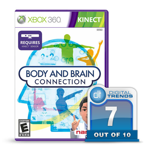
Body and Brain Connection review

Review: Namaco Bandai’s Kinect title for the Xbox 360, Body and Brain Connection, is a great idea for the family that can help create real physical and mental benefits, but suffers from a lack of variety.

Let’s be honest, the Kinect is never going to have the same types of titles that the games using controllers have. There just isn’t the same level of depth that can be utilized when you can only mimic movement and all commands are done through time based highlighting of certain areas on the TV. If you want to select something, you hold your hand over the proper icon and wait until the bar fills up and the selection registers. You will never be able to pull off a satisfying 11-hit combo that finishes with a pile driver, nor will you ever be able to effectively use cover while under fire when you don’t have the ability to walk in the direction of your choice. It just isn’t going to happen.
Instead, clever developers are not trying to recreate the standard gaming experience without hands, they are coming up with new ideas that could only work on the Kinect, and adapting popular genres that wouldn’t typically appeal to the hardcore gamers. It is a push towards the casual gaming market, as well as move to specifically target the party game crowd. And that is exactly what Namaco Bandai has done with its new title Body and Brain Connection.
The game is designed to help people by offering simple mini-games that will help to exercise your brain and keeping you physically active. Body and Brain is also a game that parents should be able to quickly get behind. In fact, it is an easy fit for the home.  If your kid likes video games, why not get them a video game designed by a “renowned neuroscientist” that can actually help them learn? The game sells itself like it is actually good for you, and it very well may be.
The idea of taking a game from the “brain training” genre is a natural fit for the Kinect.  Adding physical movements to puzzles only further expands on what can be done.  In fact, calling it a game is almost a misnomer because at its core, Body and Brain is a tool, albeit a fun one.  When you first begin the “training”, it is fun and interesting.  The more you play, the more difficult the problems become, and so the more benefit you reap from it.  But behind the good idea that is Body and Brain Connection, lies the ugly specter of repetition. Even the most enticing mini-game can grow old quickly, and while Body and Brian is an impressive title at times, it could soon wear thin on all but the most resolute of players.


How It Works
With each category, the challenges offer puzzles and problems that typically require physical actions to solve. For example in the math sections, one of the first games you will play involves simple arithmetic. The problem is displayed above, and the answer is on one of two soccer balls displayed at your avatar’s feet. Once you have the answer, you then kick the corresponding ball into the net. Another puzzle has you watching the hands on a clock before they disappear. You follow the pattern and move your arms into the proper position where the hands should be before the timer runs out. Another game features arrows displayed in various places, and you must ignore where those arrows are, and instead you move your arms in the direction that the arrows are pointing as quickly as possible.
Once you have completed the game, you can go back and replay it on a harder level of difficulty.  The arithmetic becomes algebra, the clock hands begin to move multiple spaces and disappear earlier, and the arrows appear in positions designed to try to fool you.  As you complete each game, you are awarded a grade which you can accept or attempt to approve upon.
You track your progress primarily through the “age of your brain”.  When you pop in the game for the first time, the game will run you through a few simple, time based problems. From that it will award you an age, and with each better score, and with each faster time, the age of your brain improves. Each day you will also find a new set of challenges which are offered based on previous results, in order to help you improve upon specific areas that you need help with.
The game claims to have been designed with the help of neuroscientist Dr. Ryuta Kawashima, one of the original people behind the brain-training genre, which is growing in America and is huge in Japan. The idea is that the games will help to train you mentally, while the physical nature is also beneficial to you.
The science behind it is no doubt sound, and the idea of using a video game to improve yourself is alluring. There is also a multiplayer which can make for some fun party games among people that enjoy brain-teaser based games. In that sense, Body and Brain is going right where the Kinect wants to—towards casual gamers. 
Conclusion
The idea behind this game is both fun and appealing. Play a video game and get smarter—sounds good, right? But there is also the boredom factor that will soon descend upon players of any age as you quickly burn through the existing games and puzzles. There is always the potential of more games via DLC in the future, but that is a possibility not a certainty, and even if it were guaranteed the retail package is still a bit underwhelming.
All in all, Body and Brain Connection is a great idea that will keep gamers of all ages–from kids looking to learn to adults hoping for a bit of light stimulus—entertained for a while. It is something of a Catch-22. Many that play this game every now and then will probably find it interesting and a fun distraction, but to really benefit from it you must use it constantly, which will almost certainly chase people away once the tedium begins.
The concept of this title is impressive, and hopefully more games are on the way that can turn the Xbox 360 and the Kinect into something more than just a gaming system. Body and Brain is a tool that can be used to show real and practical benefits to the lives of gamers (at least to a small degree).
Body and Brain Connection is a good game/tool for those looking for something unique to do with their Kinect, and for people that enjoy brain teasers and puzzles which could offer real life benefits to the gamer. Odds are most won’t be able to continue to play the game for too long without getting bored, but in small doses, the game is solid. If you are a Kinect owner that is looking for something worthwhile to add to your collection, then Body and Brain Connection might be the game for you.
Read More....

Brain Scientists Create Illusion of Third Human Arm

The human brain can be tricked into believing it has three arms, Swedish scientists said Thursday, giving hope to the advancement of prosthetic limbs for paralyzed stroke victims.
Scientists from the Karolinska Institute were able to convince 154 healthy volunteers that they owned multiple limbs -- to such an extent that participants broke out in a sweat when their fake limbs were threatened with a knife.
During the experiment, participants had a realistic prosthetic arm placed next to their right arm, and researchers stroked the real hand and fake hand with two small brushes in the same place. By synchronizing the strokes, the volunteers’ brains were tricked into feeling that the false arm was part of the body.
"A conflict arises in the brain concerning which of the right hands belongs to the participant's body," said researcher Dr. Arvid Guterstam. "What one could expect is that only one of the hands is experienced as one's own, presumably the real arm. But what we found, surprisingly, is that the brain solves this conflict by accepting both right hands as part of the body image, and the subjects experience having an extra third arm."
The scientists threatened either the prosthetic hand or the real hand with a kitchen knife while measuring the amount of sweating from the palm. They found results the participants had the same stress response when the prosthetic hand was threatened as when the real hand was in danger.
"It may be possible in the future to offer a stroke patient, who has become paralyzed on one side of the body, a prosthetic arm that can be used and experienced as his own, while the paralyzed arm remains within the patient's body image," said Dr. Henrik Ehrsson, who led the study.