Thursday, January 17, 2008
Mirror Neurons -- Rock Stars or Backup Singers?

Greg Hickok
Center for Cognitive Neuroscience
University of California, Irvine
Mirror neurons are the rock stars of cognitive neuroscience. Discovered in the mid-1990s by Giacomo Rizzolatti and his colleagues at the University of Parma, these brain cells have been claimed to be the neural basis for a host of complex human behaviors including imitation, action understanding, language, empathy, and mind-reading – not psychic mind-reading, but our capacity to "get inside someone else's head" and imagine how they feel or what they might do. Meanwhile, dysfunction of the mirror neuron system has been linked to developmental disorders, such as autism. With that kind of explanatory range, it's no surprise that mirror neurons have headlined in all forms of news media. But is this rock star status deserved? Will mirror neurons have the star power longevity of Mick Jagger? Or are they just back up singers?
The hidden mirror
So what exactly are mirror neurons? While studying neurons in motor areas of the frontal lobe of the Rhesus monkey brain, Rizzolatti's team noticed that some cells were responsive not only when the monkey performed an action, such as grasping a raisin, but also when the monkey simply watched the experimenter perform the same action. It was as if these neurons were simulating, or mirroring, a perceived action in the motor system of the animal. This is a very interesting and important finding, showing that sensory and motor systems interact in the brain's cortex at the single cell level.
But the interpretation of mirror neurons since then has extended well beyond sensory-motor interaction. For example, some have speculated that mirror neurons are the basis for our ability to understand the actions of others: because we know the consequences of our own actions, we can understand and anticipate the intended consequences of others' actions by activating similar neural networks in our own motor system. This concept was quickly generalized to more complex functions: because we speak, feel emotion, and have a sense of our own intentions, the theory goes, we can understand the speech of others, empathize, and "mind-read" intentions by mapping other people's behaviors onto our own mirror neuron system.
What is really being reflected?
Is the speculation that mirror neurons are responsible for "understanding" the behavior of others justified? Or are mirror neurons involved in less lofty, but nonetheless important, mental functions? A new study -- "Sensosirmotor Leaning Configures the Human Mirror System," from Current Biology (abstract or pdf download -- suggests the latter. Carolyn Catmur, Vincent Walsh, and Cecilia Heyes, researchers at University College London's Institute of Cognitive Science, stimulated the hand-related portions of motor cortex of human volunteers while they watched videos of hands performing movements of the index or little finger. Stimulation was accomplished using "transcranial magnetic stimulation" (or TMS), in which magnetic pulses are passed through the skull to induce brief electrical currents in the underlying brain tissue. TMS of motor cortex hand areas results in electrical neural impulses being transmitted to the hand itself, where these impulses can be measured by placing electrodes over the finger muscles. The researchers found that when a volunteer watched index finger movement, motor-cortex stimulation by TMS led to stronger electrical signals in the participant's own index finger compared to the pinky, and vice-versa when watching pinky finger movement. This is a mirror-neuron-like effect. Watching a video of index finger movement induces activation of the observer's own motor system controlling index finger movement. This naturally induced activity then sums with the TMS-induced activity to produce stronger than normal neural signals in the index finger muscles.
The mirror neuron theorists would say that our "understanding" of this movement is a result of this heightened activation of our own motor system. But Catmur and colleagues went beyond this basic mirror neuron result. After their initial measurements, they trained the participants to make "counter-mirror" movements: that is, when you see the index finger move, move your own pinky finger, and vice-versa. After this training, the brain responses were reassessed -- and a reversal of the mirror effect was found: watching index-finger movement resulted in more electrical activity in the pinky, and watching pinky movement produced more activity in the index finger. The brain learned new sensory-motor associations, and it is these associations that underlie the mirror neuron-like effect.
Fodder for, not parent of
This is a very nice demonstration that mirror system-like activity is subject to sensory-motor learning, suggesting it is learned rather than hard-wired. But the real question for the mirror neuron theory of action understanding is what these newly trained volunteers "understand" about these movements. Since viewing index finger movement induces activity in the participants' pinky motor systems, do they now think they are viewing little finger movement? Of course not. They still understand that they are viewing index finger movement. Conclusion: mirror system activation is not necessarily correlated with "understanding" but rather with sensory-motor learning.
This dissociation between mirror neuron-like activity and understanding comes as no real surprise. We know from decades (centuries even) of research involving patients with aphasia (language deficits resulting from brain damage, typically stroke) that it is possible to lose virtually all ability to articulate words while retaining the ability to understand the meaning of spoken words. Loss of the motor system controlling speech production, which contains the mirror system for speech, does not result in loss of the ability to understand the speech actions of others. It is also possible for the reverse situation to happen: in some patients with damage that spares the mirror system, the ability to repeat the speech of others may be intact (indicating intact sensory-motor associations), and yet they fail to understand the words. As in the study described above, mirror system function and action understanding dissociate.
The implications are clear. The mirror neuron system is not the neural basis for action understanding. This is true for simple limb actions of the sort that led to the discovery of mirror neurons in the monkey, and it is true for the first complex human behavior that the mirror neuron theory was generalized to, namely speech. If the mirror neuron theory shatters for these behaviors, its generalization to abilities like empathy or "mind-reading" seems ridiculously overstated.
This is not to say that a neural network supporting sensory-motor associations isn't important, or even that such associations are irrelevant to action understanding, language and the like. It seems quite likely that these higher-level systems make use of information derived from sensory-motor linkages. But that mirror neurons provide information that gets used by this high-level understanding does not mean that mirror neurons encode and produce this high-level understanding. You might be able to train a parrot to say "I can't get no satisfaction" -- but that doesn't mean he understands the message. Despite the hype to the contrary, mirror neurons are not the Mick Jagger of cognitive neuroscience. But there's no shame in singing backup. After all, who would want to sit through two hours of Mick singing a cappella? You need a whole band to make good music. The brain works the same way.
Gregory Hickok is professor of cognitive neuroscience and the director of the Center for Cognitive Neuroscience at the University of California, Irvine. He blogs on the neural underpinnings of language at Talking Brains and contributes to a UC Irvine cog-sci group blog as well.
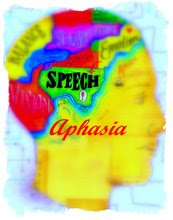
Spoken-word processing in aphasia
Wednesday, January 16, 2008
Process skill rather than motor skill ..
... seems to be a predictor of costs for rehabilitation after a stroke in working age; a longitudinal study with a 1 year follow up post discharge
Ann Bjorkdahl email and Katharina S Sunnerhagen email
BMC Health Services Research 2007, 7:209doi:10.1186/1472-6963-7-209
Published: 21 December 2007
Abstract (provisional)
Background
In recent years a number of costs of stroke studies have been conducted based on incidence or prevalence and estimating costs at a given time. As there still is a need for a deeper understanding of factors influencing these costs the aim of this study was to calculate the direct and indirect costs in a "young" (<65) sample of stroke patients and to explore factors affecting the costs.
The Mini-Mental State Examination in Behavioral Variant Frontotemporal Dementia and Primary Progressive Aphasia
How left inferior frontal cortex participates in syntactic processing: Evidence from aphasia

from Brain and Language
We report on three experiments that provide a real-time processing perspective on the poor comprehension of Broca’s aphasic patients for non-canonically structured sentences. In the first experiment we presented sentences (via a Cross Modal Lexical Priming (CMLP) paradigm) to Broca’s patients at a normal rate of speech. Unlike the pattern found with unimpaired control participants, we observed a general slowing of lexical activation and a concomitant delay in the formation of syntactic dependencies involving “moved” constituents and empty elements. Our second experiment presented these same sentences at a slower rate of speech. In this circumstance, Broca’s patients formed syntactic dependencies as soon as they were structurally licensed (again, a different pattern from that demonstrated by the unimpaired control group). The third experiment used a sentence-picture matching paradigm to chart Broca’s comprehension for non-canonically structured sentences (presented at both normal and slow rates). Here we observed significantly better scores in the slow rate condition. We discuss these findings in terms of the functional commitment of the left anterior cortical region implicated in Broca’s aphasia and conclude that this region is crucially involved in the formation of syntactically-governed dependency relations, not because it supports knowledge of syntactic dependencies, but rather because it supports the real-time implementation of these specific representations by sustaining, at the least, a lexical activation rise-time parameter.